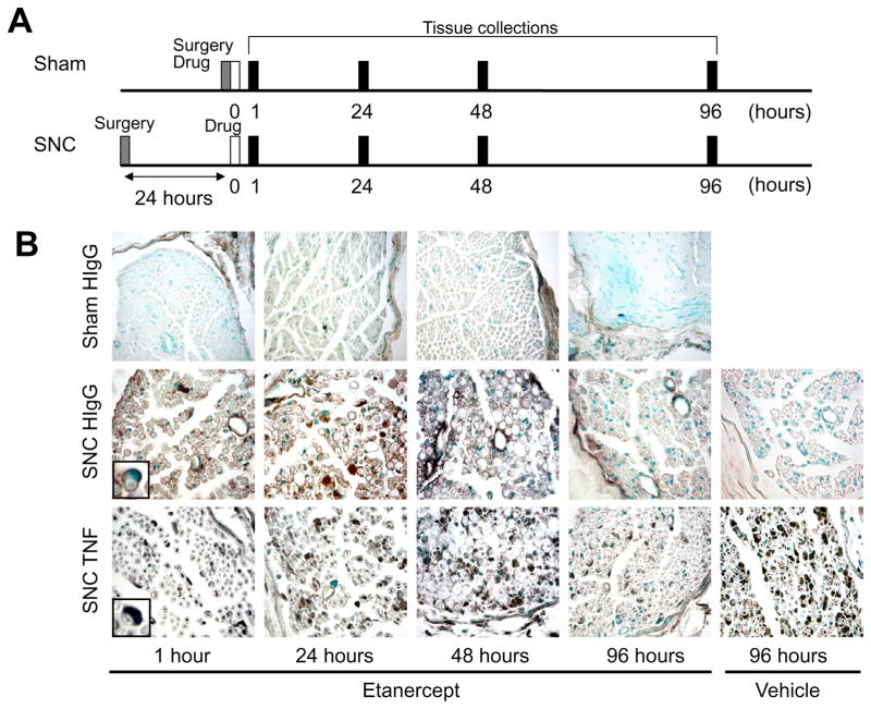
Fig. 2.
Immunohistochemistry for etanercept and TNF in nerve after the epineurial injection of etanercept. (A) Experimental schedule for surgery, therapy and tissues collection in sham and SNC groups. (B) In sham group, etanercept immunoreactivity using anti-human IgG (HIgG, brown) was observed in the epineurial space and the perineurium at 1–96 h after its injection. It was not detectable in the endoneurial space at either time point. In contrast, etanercept reached the endoneurial space after its epineurial injection to the injured rat sciatic nerve. Representative micrographs of n=8 –10 rats/group. Insets are magnified images of myelinated axons with surrounding Schwann cell cytoplasm. Magnification 200× (Sham). Magnification 400× (SNC).

