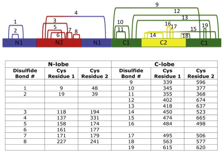
Figure 2.
Disulfide bonds of hTF. The 19 disulfide bonds formed within hTF are shown numbered according to primary sequence. Equivalent disulfide bonds from each lobe are listed on the same line. Individual subdomains are colored accordingly: N1, blue; N2, red; C1, green; C2, yellow. See the color plate.

