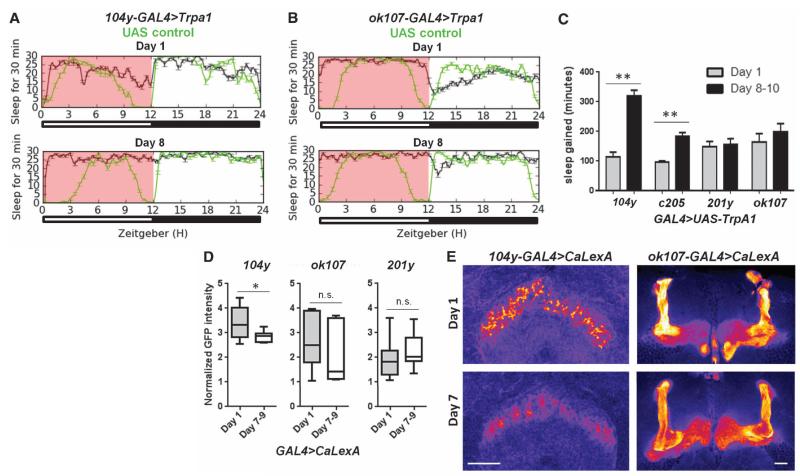
Fig. 4. The sleep-promoting dFSB is more active in young flies.
(A and B) Sleep traces over 24 hours in GAL4>UAS-dTrpA1 flies (black) or UAS controls (green) at different ages. The pink box denotes elevated temperature. The y axis denotes sleep episodes per 30 min. (C) Quantification of sleep gained during 12 hours of elevated temperature (pink box) in sleeppromoting GAL4>UAS-dTrpA1 lines at multiple developmental time points compared to UAS controls (from left to right, n = 10, 6, 12, 12, 12, 12, 10, and 8 flies, with multiple independent replicates). (D) Normalized GFP intensity of the indicated brain region in GAL4>UAS-CaLexA flies of different ages (104y-GAL4: n = 9 day-1, n = 10 day-7 to -9; ok107-GAL4: n = 15 day-1, n = 14 day-7 to -9; 201y-GAL4: n = 22 day-1, n = 16 day-7 to -9). (E) Representative images of the dFSB (left) and MB (right) in brains immunostained for GFP. GFP is pseudocolored fire. Scale bars, 37.5 μm. **P < 0.0001, *P < 0.05; one-way ANOVA with Tukey’s post-hoc test (C); and unpaired two-tailed Student’s t test plus Welch’s correction (D).

