Fig. 6.
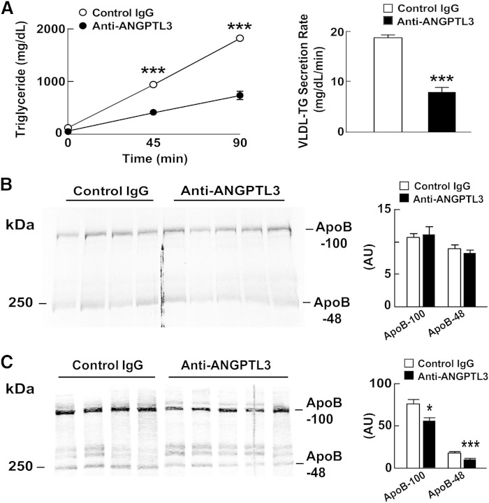
ApoB secretion in REGN1500-treated WT mice. A: WT mice were synchronized for 3 days (fasting: 5:00 PM to 7:00 AM and feeding: 7:00 AM to 5:00 PM). On day 4, mice were refed at 7:00 AM and injected at 9:00 AM with either a control antibody or REGN1500 (10 mg/kg) (n = 6 male mice per group, 10 weeks old). Two hours after the injection, Triton WR1339 (500 mg/kg) and 200 μCi of [35S]methionine were injected into the tail vein. Blood samples were drawn from the tail veins before and 45 min after the injection. The mice were euthanized at 90 min and blood was collected. Plasma TG levels were measured at the indicated time after Triton WR1339 injection. B: Plasma collected at the 90 min time point was delipidated and size fractionated by SDS-PAGE (5%). Gels were dried and exposed to X-ray film (BIOMAX XAR; Kodak, catalog number 1651579) for 4 days at −80°C. The films were scanned using a HP Scanjet 5590 and quantified using ImageJ. The intensity of each band was corrected for background using a blank from the same film. C: The same plasma samples were subjected to immunoblot analysis with an anti-ApoB antibody and the bands were quantified using an Odyssey Image Analyzer (Li-Cor). *P < 0.05, ***P < 0.001.

