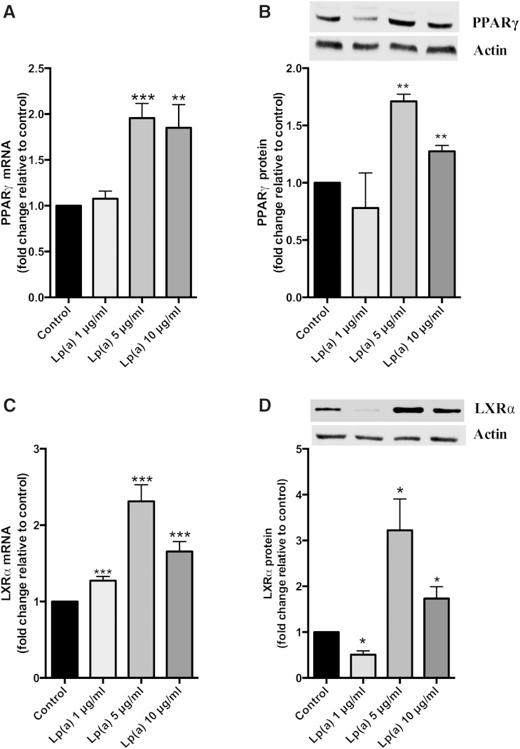
Fig. 2.
Lp(a) stimulates PPARγ-LXRα expression. HepG2 cells were treated with 1, 5, and 10 μg/ml purified Lp(a) protein for 12 h at 37°C. A: PPARγ mRNA levels as determined by RT-PCR. PPARγ mRNA was normalized to β2-microglobulin and GAPDH mRNA levels and expressed relative to those of the control untreated cells. B: PPARγ protein levels as determined by Western blot. PPARγ protein levels were normalized against actin (inset) and expressed relative to those of untreated cells. C: LXRα mRNA levels as determined by RT-PCR normalized to β2-microglobulin and GAPDH and expressed relative to control. D: LXRα protein levels normalized to actin and expressed relative to control. Results are expressed as mean ± SE for two experiments performed in triplicate for RT-PCR and triplicate Western blots for protein quantification. *P < 0.05, **P < 0.01, ***P < 0.001 compared with control.

