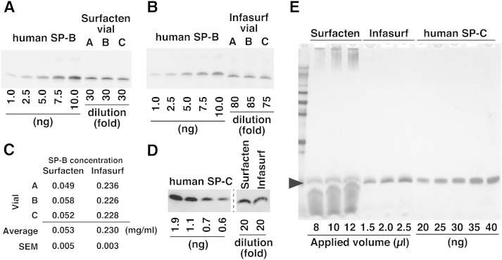
Fig. 1.
Analysis of SP-B and SP-C by Western blotting. A, B: Western blot analysis of SP-B in Surfacten (A) and Infasurf (B). Each sample lane contains 2.5 μl of diluted surfactant drug. C: Quantification of SP-B based on Western blotting. Data for each vial is calculated from three experiments. D: Western blot analysis of SP-C in Surfacten and Infasurf. Each sample lane contains 9 μl of diluted drug. E: Silver staining of gels after SDS-PAGE reveals that some materials comigrate with SP-C (arrowhead) in surfactant drugs. Proteins are also seen in the upper part of the gel in Surfacten lanes.

