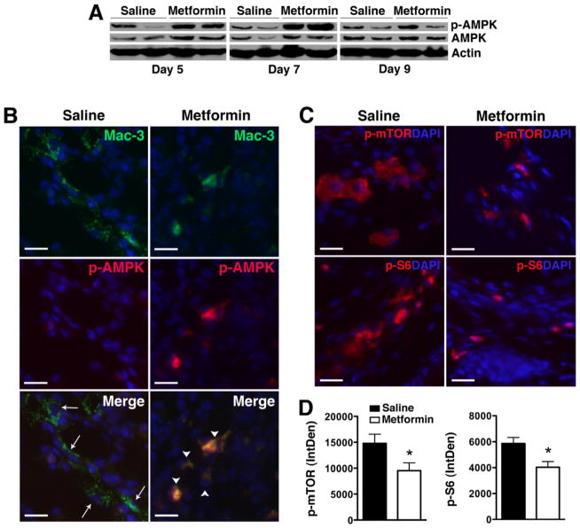
Figure 4. Metformin activates AMPK and suppresses mTORC1 activity in KRN arthritis.
(A) Protein lysates from day 5, 7, or 9 paws obtained from KRN arthritic mice were probed for p-AMPK and total AMPK. Actin served as control for protein loading. (B) Day 9 paws were stained for Mac-3 (arrows, green) and p-AMPK (red). Colocalization (arrowheads) appeared orange/yellow. (C) Day 9 paws were stained for phospho (p)-mTOR and p-S6 (red). DAPI (blue) stained nuclei. Scale bar = 25 μm. (D) Intracellular level of p-mTOR and p-S6 was analyzed using ImageJ program as detailed in the Materials and Methods section and presented as integrated optical density (IntDen). Values represent mean ± SEM, n = 4–5 mice per treatment group. *P < 0.05 compared with saline.

