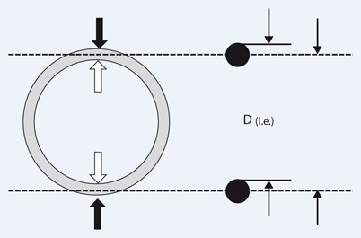
Fig. 2.

Measurement according to the outer-to-outer edge (black arrow), the inner-to-inner edge (white arrow), and the leading-edge method (right): outer wall reflection–inner wall reflection [D(l.e.)] of the opposing aortic wall in order to minimize and standardize the ultrasound overestimation of vessel thickness (black dots) caused by the blooming effect at boundaries with high acoustic impedance mismatches (such as vascular wall/blood). (Modified from [20])
