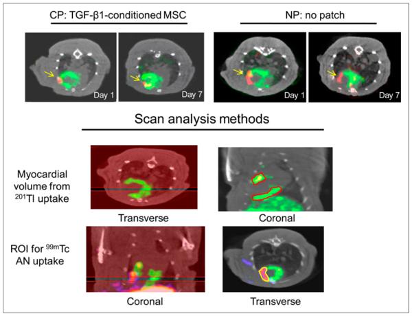
FIGURE 2.
Noninvasive in vivo imaging. (Top panel) Transverse image from dual-Risotope SPECT/CT scans from representative experiments from CP and NP groups that were injected with 201Tl and 99mTc-annexin V and imaged at days 1 and 7 after LAD ligation. 201Tl uptake in perfused myocardium is shown in green and focal uptake of 99mTc-annexin V in red, with arrows pointing to this uptake. CP-treated rats showed greater decrease in 99mTc-annexin V in infarcted area between days 1 and 7 than did NP-treated rat. (Lower panel) Methods for quantification of scan data using InVivo-Scope software. Regions are drawn around 201Tl uptake on sequential 5-slice coronal sections from septum to lateral wall and myocardial volume measured using volume calibration. Similarly, regions are drawn around 99mTc uptake on sequential 4-slice sections and tracer uptake measured as millicuries, which is divided by decay-corrected ID for %ID. AN = annexin.

