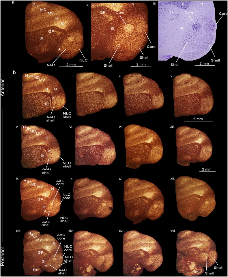
Fig 2. PVALB mRNA expression in the core and shell regions of the NLC and AAC song nuclei in the budgerigar brain.
(a) High power view of darkfield (i-ii) and brightfield (cresyl violet stained) (iii) images showing expression of parvalbumin mRNA. (b) Serial sections of a budgerigar brain (i-xvi) showing the core and shell regions of NLC and AAC song nuclei. Silver grains is white; cresyl violet is red-brown. Sections are in the coronal plane; medial is to the left, dorsal is top. See list for abbreviations.

