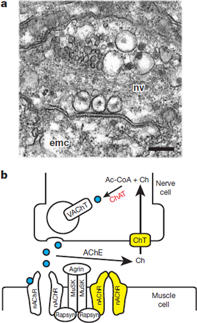
Figure 2. The neuromuscular junction in Hydra.
a, Electron micrograph of a nerve synapsing on a Hydra epitheliomuscular cell. emc, epitheliomuscular cell; nv, nerve cell. Three vesicles are located in the nerve cell at the site of contact with the epitheliomuscular cell. Scale bar, 200 nm. b, Schematic diagram of a canonical neuromuscular junction. Yellow indicates presence in Hydra. Choline acetyltransferase (ChAT) is shown in red because it is not clear whether Hydra has an enzyme that prefers choline (Ch) as a substrate. Acetylcholine (ACh) molecules are shown as blue circles. The nicotinic acetylcholine receptor (nAChR) is shown in the open state with acetylcholine bound (left), and in the closed state in the absence of bound acetylcholine (right). AChE, acetylcholinesterase; ChT, choline transporter; MuSK, muscle-specific kinase; VAChT, vesicular acetylcholine transporter.

