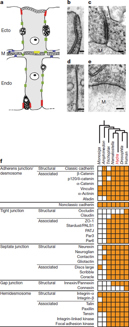
Figure 3. Hydra cell junctions.
a, Schematic diagram of the positions of cell–cell and cell–matrix contacts in Hydra epitheliomuscular cells. Septate junction, red; gap junctions, green; spot desmosomes, blue; hemidesmosome-like cell–matrix contact, yellow. Ecto, ectodermal cell; Endo, endodermal cell; M, mesoglea. For simplicity the nervous system has been omitted. b–e, Electron micrographs of cell–cell and cell–matrix contacts in Hydra. b, Apical septate junction. c, Spot desmosome between basal muscle processes. d, Gap junction in the lateral cell membrane. e, Hemidesmosome-like cell–mesoglea contact site. Scale bars in b–e indicate 100 nm. f, Phylogenetic distribution of cell–cell and cell–substrate contact proteins. A filled box indicates the presence of an orthologue from the corresponding protein family as identified by SMART/Pfam analysis or conserved cysteine patterns. See Supplementary Information section 17 and Supplementary Table 21 for details.

