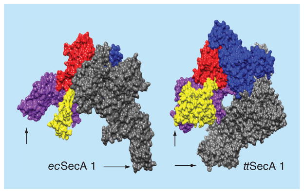
Figure 2. Space filling models of SecA dimers.

Dimeric SecA proteins were structurally aligned on one of their protomers (the nucleotide binding domain NBD, red; the intramolecular regulator of ATPase 2 IRA2, dark blue; the protein binding domain PBD, yellow and the C-domain, purple) so as to demonstrate the variable position that the second (grey) protomer occupies; arrows indicate C-terminus. The figure was created using the UCSF Chimera package [67,68]. The structures used are: Escherichia coli (ecSecA1; 2FSF) [51]. Thermus thermophilus (ttSecA; 2IPC) [61].
For color images please see online www.future-science.com/doi/full/10.4155/FMC.15.42
