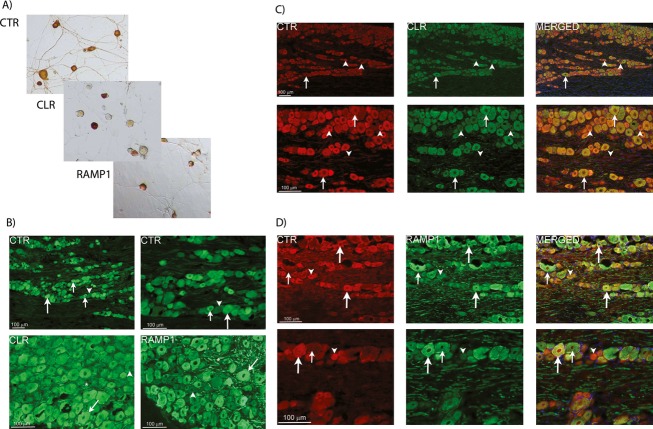
Figure 1.
Immunostaining of CGRP-responsive receptor components in rat TG. (A) Expression of CLR, CTR, and RAMP1 (CGRP and AMY1 receptor components) in isolated TG neurons by immunostaining. Images are representative of those obtained in at least three independent preparations. (B) Expression of CGRP receptor components in rat TG. CTR is expressed in small to medium sized cells (small arrows) and some larger neurons (large arrows). CLR and RAMP1 are expressed mostly in larger neurons (arrows) and thick nerve fibers (asterisks). Arrowheads indicate negative neurons. (C) Double-staining of CTR and CLR in rat TG. No clear co-expression is found between CTR (red) and CLR (green) in the TG neurons. CTR-positive neurons and fibers (arrowheads) lacking CLR expression are found. Arrows point at neurons only expressing CLR. DAPI, (blue) staining nuclei, is used in the merged picture. (D) Double-staining of CTR and RAMP1 in rat TG. Large arrows indicate CTR and RAMP1-positive neurons and their co-localization in the merged pictures. Large neurons only expressing RAMP1 are found (small arrows). Neurons only expressing CTR are also found (arrowheads). DAPI, (blue) staining nuclei, is used in the merged pictures. For (B–D), data are representative images from four rats. CGRP, calcitonin gene-related peptide; TG, trigeminal ganglia; CLR, calcitonin receptor-like receptor; CTR, calcitonin receptor; RAMP1, receptor activity-modifying protein 1; DAPI, 4’6-diamidino-phenylindole.

