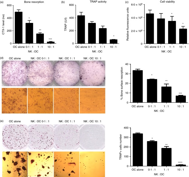
Figure 3.
The killing of osteoclasts by interleukin-15 (IL-15) -activated natural killer (NK) cells is ratio-dependent. Enriched osteoclasts (5 × 10⁴/well) were seeded in a 96-well plate on bone slices or plastic and co-cultured with IL-15-activated NK cells, at effector : target ratios of 0.1 : 1, 1 : 1 and 10 : 1, for 3 days in the presence of macrophage colony-stimulating factor (M-CSF; 25 ng/ml) and receptor activator of necrosis factor κB ligand (RANKL; 10 ng/ml). Supernatants were collected for measurements of: (a) Osteoclast-mediated collagen type I degradation (C-terminal type I collagen fragments; CTX-I) and (b) tartrate-resistant acid phosphatase (TRAP) activity. (c) At the end of the culture, cells were incubated with 10% Presto Blue to determine cell viability. The mean ± SEM values are shown for n = 4 donors. (d) Bone slices were removed from wells, washed and stained with haematoxylin, to enable visualization of the pits resorbed by osteoclasts. An Immunospot Image Analyser was used to quantify the resorbed bone pits on the surface of the bone slice, indicated by the darker areas (top panel). The pits were also visualized under a microscope (bottom panel, magnification × 100). (e) The osteoclasts seeded on plastic were fixed and TRAP stained, and the number of TRAP+ osteoclasts was quantified using an Immunospot Image Analyser (top panel) as described. Magnification × 50 (bottom panel). Figures are representative of n = 4 donors. *P < 0·05; **P < 0·01; ***P < 0·001; ****P < 0·0001.

