
Figure 4.
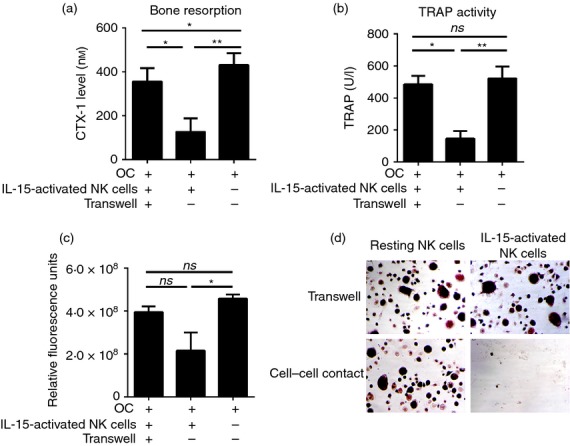
Cell–cell contact is required for the lysis of osteoclasts by interleukin-15 (IL-15) -activated natural killer (NK) cells. NK cells were isolated from peripheral blood mononuclear cells and cultured in the presence or absence of IL-15 as described. Osteoclasts were differentiated and harvested as described. Enriched osteoclasts (5 × 10⁴/well) were seeded into the lower chamber of a 96-well transwell on bone slices or plastic. On the following day, 2·5 × 105 IL-15-activated NK cells or resting NK cells were added into either the upper chamber (transwell) or together with osteoclasts into the lower chamber, and co-cultured for 3 days with macrophage colony-stimulating factor (M-CSF; 25 ng/ml) and receptor activator of necrosis factor κB ligand (RANKL; 10 ng/ml). The culture supernatants were collected for detection of: (a) osteoclast-mediated collagen type I degradation (C-terminal type I collagen fragments; CTX-I) and (b) tartrate-resistant acid phosphatase (TRAP) activity. (c) Cells were incubated with Presto Blue reagent to determine the viability of the cells. The mean ± SEM values of three independent experiments are shown for n = 6 donors. (d) Adherent osteoclasts were fixed with 4% paraformaldehyde and TRAP stained. Magnification × 50. Figures are representative of n = 6 donors. ns, not significant; *P < 0·05; **P < 0·01.