
Figure 2.
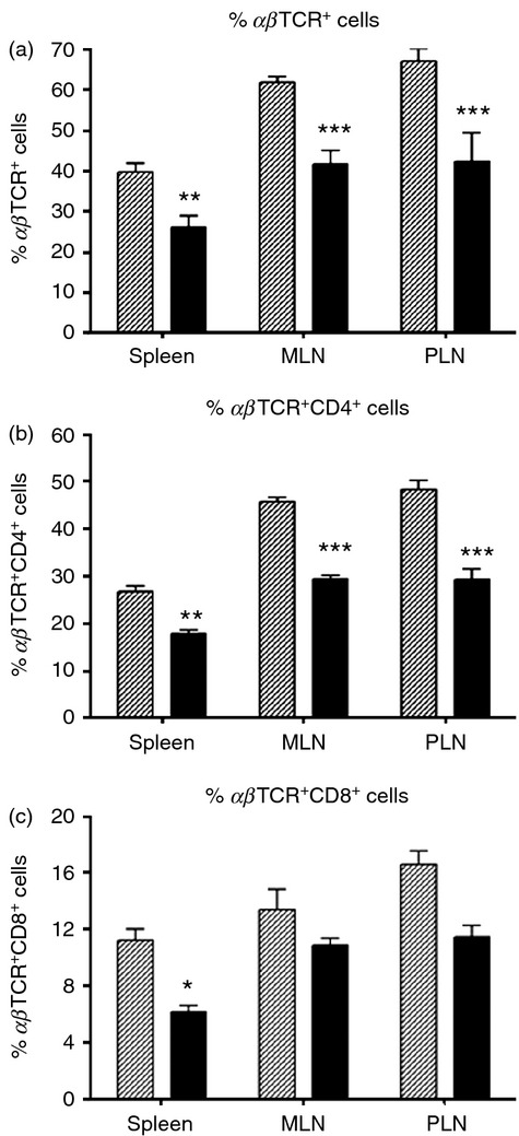
T-cell deficits in diabetic LEW.1AR1-iddm rat spleen, mesenteric lymph node (MLN) and pancreatic lymph node (PLN). (a) Diabetic LEW.1AR1-iddm rats (solid bars, n = 5–6) had fewer αβTCR+ cells compared with control LEW.1AR1 rats (hatched bars, n = 7 or 8) in all three tissues; (b) diabetic rats were also deficient in αβTCR+ CD4+ cells and (c) a significant decrease in αβTCR+ CD8+ cells was observed in the spleen. Data are mean ± SD, analysis of variance followed by Scheffé post-hoc test. *P < 0·05, **P < 0·01, ***P < 0·001.