Figure 1.
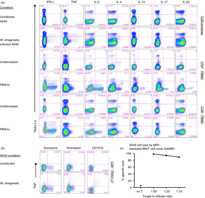
Mycobacteria reactive CD8+ TRAV1–2+ cells produce interferon-γ (IFN-γ), tumour necrosis factor (TNF), and are cytotoxic. (a) Representative dot plots of T-cell responses from the cytokine-staining assay are shown. CD8+ T cells enriched from peripheral blood mononuclear cells (PBMC) were incubated overnight with A549 stimulator cells left uninfected or infected with Mycobacterium smegmatis (rows 1 and 2). Alternatively, PBMC were stimulated for 6 hr with PMA/ionomycin (PMA/io). A protein transport inhibitor was added for the last 6 hr of the assay to prevent cytokine release except for the detection of interleukin-10 (IL-10) and TNF. Cells were surface stained for the expression of TRAV1–2, CD8, CD4, and then fixed and permeabilized before staining for cytokines. Cytokines are listed on the x-axis and TRAV1–2 on the y-axis. The numbers represent the frequency of events from the CD8+ gate (rows 1, 2, 5 and 6) or CD4+ gate (rows 3 and 4). Similar responses were detected from a minimum of three individuals. (b) Representative dot plots of CD8-enriched T cells from PBMC in response to uninfected or M. smegmatis infected A549 are shown. The numbers represent the frequency of events from CD8+ TRAV1–2+ gate. The y-axis represents expression of TNF and the x-axis expression of granzyme B, granulysin, or CD107a. Data are representative of a minimum of three individuals. (c) The graph depicts the per cent specific lysis of M. smegmatis-infected A549 cells by MHC-like molecule-1 (MR1) -restricted mucosa-associated invariant T (MAIT) cell clone D426B1 at different target to effector ratios (1 : 50, 1 : 25 and 1 : 10). No target cell lysis was observed in the absence of infection.

