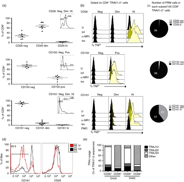
Figure 3.
CD26, CD150 and CD161 specifically identify functional MHC-like molecule-1 (MR1) -restricted mucosa-associated invariant T (MAIT) cells. (a) Frequency of CD8+ cells that express CD26 (neg, dim and high), CD150 (neg and pos) and CD161 (neg, dim and high) cells (n = 6) in the absence of stimulation. The insets depict the expression levels of CD26, CD150 or CD161 from CD8+ cells and represent the gating strategy used for the sorted subsets tested in (b). (b) Overlay histograms depicting the expression of tumour necrosis factor (TNF) by CD8+ TRAV1–2+ events from each sorted cell population (CD26 neg, dim, hi) (CD150 neg, pos) (CD161 neg, dim, hi) under each condition. Sorted cell populations were rested in medium overnight (37°, 5% CO2) before overnight incubation with uninfected or Mycobacterium smegmatis-infected A549 cells in the presence of α-MR1 (clone 26.5) blocking or control (msIgG2a) antibodies. The following day, the cells were stained and analysed as described in the Methods. No TNF was detected by CD8+ TRAV1–2+ cells in the CD26dim subset or by cells lacking CD26, CD150 or CD161 in response to stimulation. The fluorescence minus one (FMO) condition represents the background level of staining observed in the absence of TNF-α antibody under conditions with infected antigen-presenting cells. Similar results were obtained using peripheral blood mononuclear cells from three different individuals. (c) Average (n = 3) number of CD8+ TRAV1–2+ TNF+ cells that express CD26hi (99·2 ± 0·4), CD150hi (97·5 ± 1·8), CD161dim (43 ± 7·3), or CD161hi (56·9 ± 7·4) per 100 CD8+ TRAV1–2+ cells based on results from (a) and (b). (d) Histograms representing CD161 (left) and CD26 (right) expression on FACS-sorted CD8+ TRAV1–2+ CD26hi in response to uninfected and M. smegmatis-infected A549 cells. The frequency of cells lacking CD161hi expression after stimulation with infected cells is depicted in the left histogram. This result was representative of three biological replicates where on average 77% of stimulated cells lacked CD161hi expression. (e). TRAJ gene usage associated with mucosa-associated invariant T MAIT cells, namely TRAJ12, TRAJ20 and TRAJ33, from TRAV1–2 sequences determined from FACS-sorted CD8+ TRAV1–2+ cells expressing CD26hi or lacking CD26 from two individuals (D433 and D462). ‘Other’ represents all non-TRAJ12, TRAJ20 and TRAJ33 genes used.

