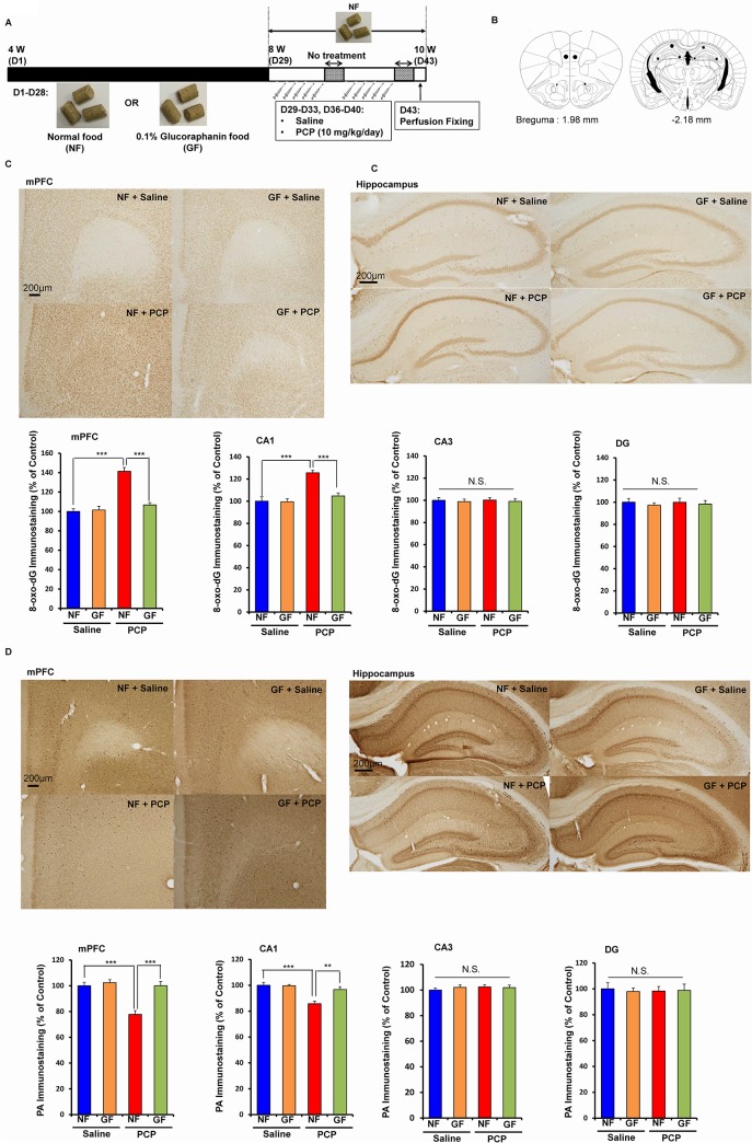
Fig 5. Prophylactic effect of dietary GF food during juvenile and adolescence on PCP-induced oxidative stress and reduction of PV-positive cells in the brain at adulthood.
(A): Schedule of treatment, and immunohistochemistry. From days 1 (4-week olds)– 28 (8-week olds), normal food (NF) or 0.1% glucoraphanine food (GF) was administered into mice. From days 29 (8-week olds), normal food was administered to all mice. Subsequently, saline (10 ml/kg/day) or PCP (10 mg/kg/day) was administered from days 29–33 and days 36–40. On day 43, all mice were perfused. (B): Brain regions of mPFC, CA1, CA3, and dentate gyrus (DG) of hippocampus, were shown. (C): Repeated PCP administration significantly increased 8-oxo-dG immunostaining in the mPFC and CA1, but not CA3 and DG. Dietary intake of 0.1% GF from 4-week old to 8-week olds significantly attenuated PCP-induced increases of 8-oxo-dG immunostaining in the mPFC and CA1 at 10-week olds. The data show the mean ± SEM (n = 5–7). (D): Repeated PCP administration significantly decreased PV-positive immunostaining in the mPFC and CA1, but not CA3 and DG. Dietary intake of 0.1% GF from 4-week old to 8-week olds significantly attenuated PCP-induced decreases of PV-positive immunostaining in the mPFC and CA1 at 10-week olds. The data show the mean ± SEM (n = 5–7). **P < 0.01, ***P < 0.001, N.S. not significant.

