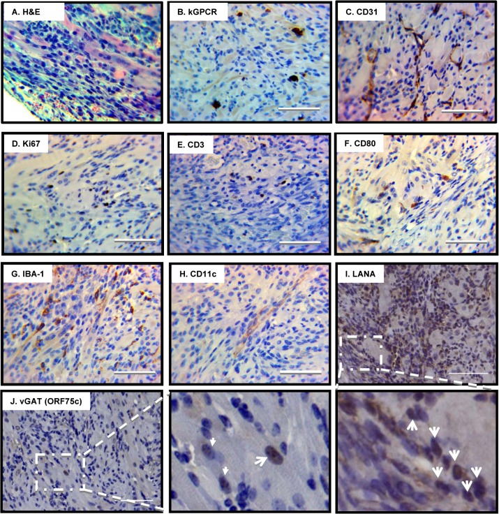
Fig 6. Pathological analysis of angiogenic tumors derived from γHV68.kGPCR-infected mice.
Subcutaneous tumors developed in γHV68.kGPCR-infected BALB/c mice were analyzed by hematoxylin & eosin (H&E) staining (A) and immunohistochemistry staining with antibodies against the HA epitope (kGPCR, B), endothelial marker CD31 (C), proliferation marker Ki67 (D), T cell marker CD3 (E), CD80 (F), macrophage marker IBA-1 (G) and dendritic cell marker CD11c (H). Tumors were also analyzed by immunohistochemistry staining with antibodies against γHV68 LANA (ORF72, latent antigen) (I) and vGAT (ORF75c, lytic antigen) (J). Boxed regions were amplified right below (I) or next (J) to the original images. Scale bars denote 25 μm.

