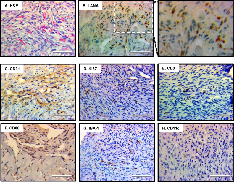
Fig 7. Angiogenic and inflammatory features of human Kaposi’s sarcoma.
Human Kaposi’s sarcoma lesion was analyzed by H&E staining (A) and immunohistochemistry staining with antibodies against the latent nuclear antigen LANA (B), markers for endothelial cells CD31 (C), proliferating cells Ki67 (D), T cells CD3 (E), antigen-presenting cells CD80 (F), macrophage IBA-1 (G) and dendritic cells CD11c (H). Representative images were shown and scale bars denote 25 μm.

