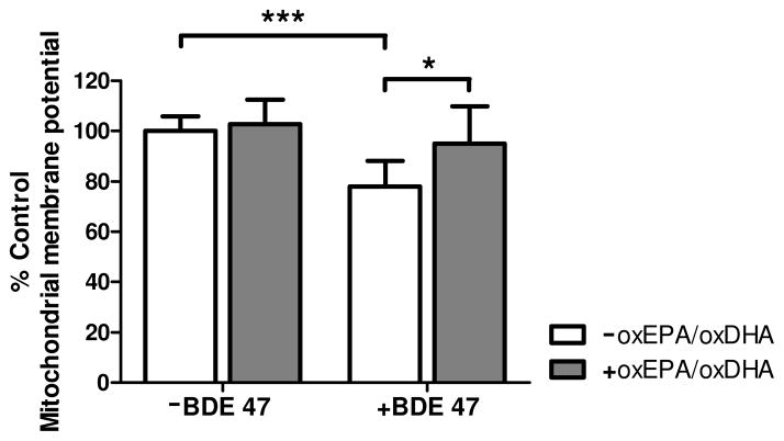
Figure 4. Effect of oxEPA/oxDHA on loss of mitochondrial membrane potential from BDE 47 exposure.
Mitochondrial membrane potential (ΔΨm) was quantified in cells by measuring JC-fluorescence in cells pretreated with, or in the absence of oxEPA/oxDHA for 12 h, followed by 24 h exposure to 100 μM BDE 47 (or DMSO). *p≤0.05, ***p≤0.001 relative to the corresponding control group. Data are expressed as percent of control values and are means ± SEM of n=3 experiments.

