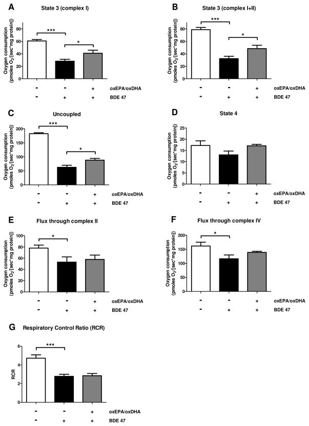
Figure 5. Effect of oxEPA/oxDHA on loss of mitochondrial electron transport system function from BDE 47 exposure.
Oxygen consumption (flux) was determined in permeabilized HepG2 cells exposed to: 1) vehicle control (DMSO, 36 h), 2) DMSO for 12 h followed by BDE 47 (100 μM) for 24 h, or 3) pretreated with oxEPA/oxDHA for 12 h before BDE 47 exposure for 24 h. Experimental conditions included: (A) State 3 respiration with complex I substrates only; (B) maximally-induced State 3 respiration with substrates of complex I and II; (C) maximum uncoupled respiration using the uncoupling agent CCCP; (D) State 4 respiration induced by oligomycin. Oxygen flux capacity through (E) complex II, and (F) complex IV were also determined. (G) The respiratory control ratios (RCRs) were calculated as the ratio of maximally-induced State 3 respiration/State 4 respiration. *p≤0.05, ***p≤0.001 relative to control group. All data are means ± SEM of n=4 experiments.

