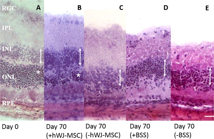
Fig 1. Histology of experimental groups.
A representative histology at day 0 (A) and day 70 (B-E) of different experimental groups. At day 70, the ONL were clearly detectable in eyes treated with hWJ-MSCs (B) compared to a thin layer or almost absent ONL in contralateral non-injected eye (C). The ONL was undetectable in the BBS injected eye (D) or the follow control eye (E). RGC: retinal ganglion cell, IPL: inner plexiform layer, INL: inner nuclear layer (indicated by double-headed arrows), ONL: outer nuclear layer (indicated by asterisks), RPE: retinal pigment epithelium. Scale bar represents 20μm.

