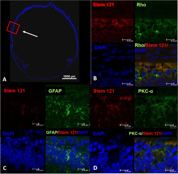
Fig 5. Confocal microscopy of the whole eye injected with hWJ-MSCs.
Confocal microscopy of the whole eye (A) and magnified images of the injected site (B-D). Red box represents the magnified area and the white arrow indicates the injection site. Co-localisation of DAPI (blue) and stem 121 (red) with rhodopsin (green), GFAP (green) and PKC-α (green) was detected at day 70 post injection, indicating that hWJ-MSCs has the potential to differentiate to retinal neuronal cells. Scale bar represents 10 μm.

