
Fig 3. Proinsulin is dislocated into the cytosol and degraded by the proteasome.
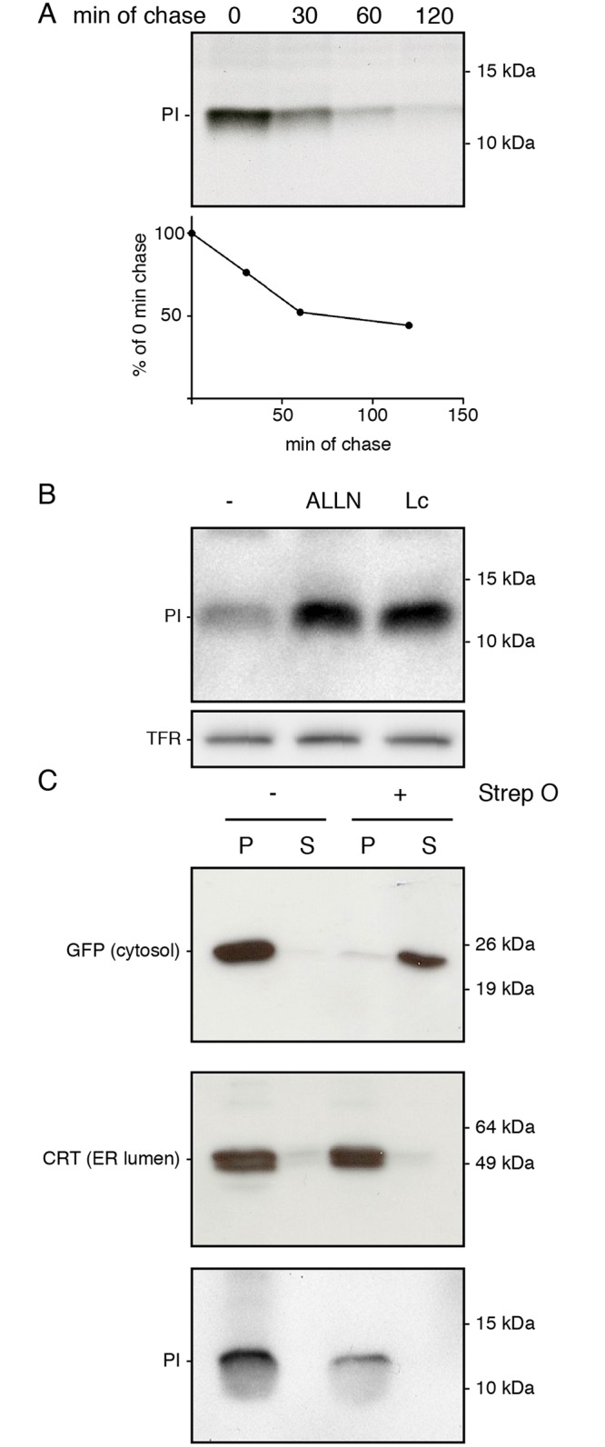
(A) K562 cells stably expressing preproinsulin were pulse labeled with 35S-methionine and cysteine for 15 minutes and chased for the indicated times. Immunoprecipitated proinsulin was analyzed using 15% SDS-PAGE. Quantification of the pulse chase experiment is shown below. Gel is representative for four experiments. (B) Preproinsulin-expressing K562 cells were treated for 3 hours with either DMSO, 100 μM ALLN or 10 μM Lactacystin (Lc). Cells were lysed and proteins were separated using 12% Nu-PAGE. Proinsulin levels were analyzed by Western blot. Transferrin receptor (TFR) was blotted as a loading control. Gel is representative for four experiments. (C) K562 cells stably expressing preproinsulin were treated for 3 hours with 100 μM ALLN and treated with Streptolysin-O to permeabilize the plasma membrane. After separation of the cytosol (supernatant) from the cell (pellet), protein levels for GFP (top panel), calreticulin (CRT, middle panel) and proinsulin (PI) were analyzed using 15% SDS-PAGE and Western blot. Gels are representative for three experiments.