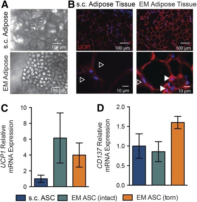
Figure 1.
Morphology and expression profiles of EM adipose biopsies resemble human brown/beige fat. (A): Low-magnification light microscope images of freshly isolated biopsies of EM and s.c. adipose tissues. (B): Histological staining of adipose tissue sections for the brown fat marker UCP1. Closed versus open arrowheads indicate multilocular versus unilocular adipocytes. Quantitative polymerase chain reaction expression of the brown fat gene UCP1 (C) and the beige gene CD137 (D) relative to patient-matched s.c. biopsies for the indicated EM adipose biopsies as a function of rotator cuff tear state. Abbreviations: ASC, adipose-derived stem cell; EM, epimuscular.

