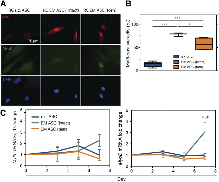
Figure 5.
EM ASCs exhibit increased stiffness-directed early myogenesis. (A): Immunofluorescent images of ASCs cultured on hydrogels of muscle-like stiffness and stained for two transcriptional markers of myogenesis, Myf5 (top) and MyoD (middle). (B): Quantification of Myf5 immunofluorescence in these cultures. A positive signal was determined relative to an undifferentiated control for each culture. ∗, p < .05; ∗∗, p < .01; ∗∗∗, p < .001. (C): Gene expression data for Myf5 and MyoD at day 7 for the indicated sources and tear states. ∗, p < .05 compared with RC s.c. ASCs; #, p < .05 compared with RC EM ASCs (torn). Abbreviations: ASCs, adipose-derived stem cells; DAPI, 4′,6-diamidino-2-phenylindole; EM, epimuscular; RC, rotator cuff.

