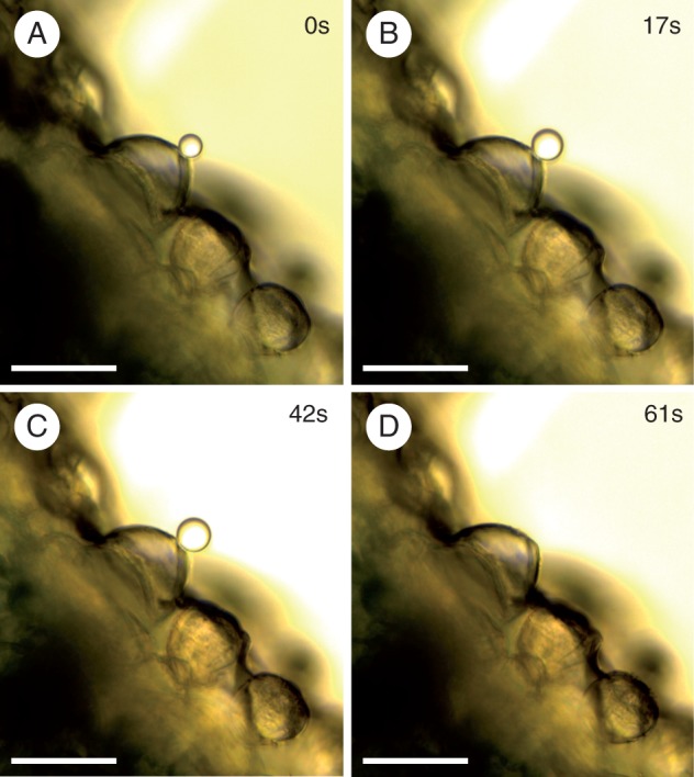
Fig. 2.

Micrographs showing secretion from sessile hydathode trichomes on the abaxial leaf surface of Rhinanthus alectorolophus. The secretion was observed in oil shortly after immersion of the sample (0s, A) and in the time series as indicated (B–D). The drop of liquid finally detached from the trichome and moved out of view (D). The scale bars indicate 50 μm.
