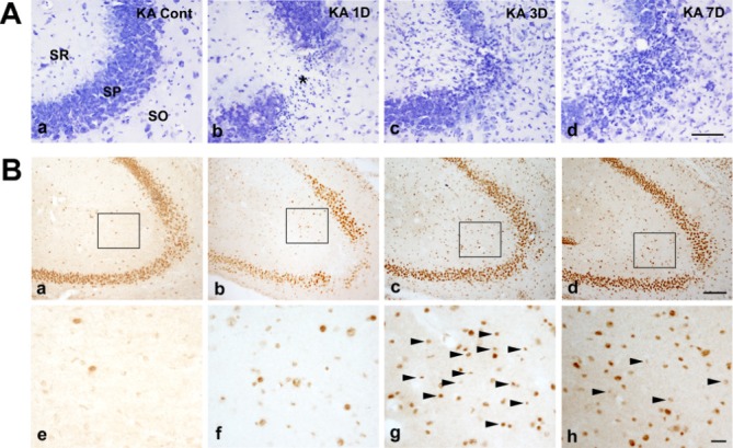
Fig. 1. Representative images with Cresyl violet staining and immunohistochemical staining with anti-GDF15 primary antibodies in kainic acid (KA) treated ICR mice. (A) In the KA-group, cresyl violet-positive cells were decreased in the striatum pyramidal (SP) of the hippocampal CA3 region (asterisk in Ab). In addition to pyramidal cell loss, small glial cell immunoreactivity was evident (Ac, d). (B) GDF15 immunohistochemistry in the hippocampal CA3 region of the control and KA groups at 1, 3 and 7 days after KA injection. GDF15 immunoreactivity was markedly increased in the KA group (Aa~d). Higher magnification images of the rectangular area Aa-d in the hippocampus show sequential changes in the GDF15 expression (Ae~h). GDF15 immunoreactivity was found in relative small nuclei than those in control (arrowheads). SO, stratum oriens; SP, stratum pyramidale; SR, stratum radiatum. Scale bars=100 µm in A, 200 µm in Ba~d, 20 µm in Be~h.

