. 2015 Jun 17;24(2):146–155. doi: 10.5607/en.2015.24.2.146

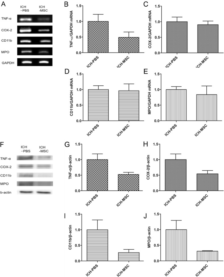
Fig. 4. RT-PCR and western blot analysis of inflammatory factors. The mRNA and protein levels were detected in whole brain tissue of ICH rats at 3 days after transplantation. (A) mRNA levels of inflammatory factors (TNF-α, COX-2, CD11b, and MPO) by RT-PCR. (B~E) Quantification of the mRNA expression of inflammatory factors in the hUCB-MSCs-treated group compared with the controls. (F) Representative western blots of the four inflammatory factors (G~J). Quantification (protein expression) of inflammatory factors in the hUCB-MSCs-treated group compared with the controls. mRNA and protein levels of inflammatory factors were lower in the hUCB-MSCs-treated group than in the controls (not significant). Densitometric analysis was performed using the Image J software by normalizing to GAPDH for RT-PCR and β-actin for western blot as the loading control; n=3 per group.

Fig. 4