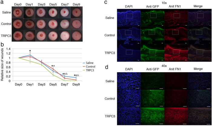
Figure 7. Wound contraction was enhanced by the transplantation of TRPC3 overexpressing fibroblasts in mice.
To assess the effect of TRPC3 overexpressing fibroblasts on wound healing in vivo, TRPC3 overexpressing fibroblasts, empty-vector transfected control fibroblasts or the same amount of saline were injected into the dorsum of nude mice. 10 days post-transplantation, 6 mm wounds were created and the size of the wounds were subsequently measured on Days 1, 3, 5, 7 and 9. The wound tissue was ultimately harvested at Day 9 post-wounding and immunochemistry with anti-TRPC3 and Fibronectin was performed. a; Representative photographs of cutaneous wounds. Differential healing was observed at all time points checked (n = 6 for each time point). b; Quantification of the wound area from Days 0–9. The data is expressed as the mean ± SD. n = 6 per group. *p < 0.05 TRPC3 versus Saline. p < 0.05 TRPC3 versus PMXS. p < 0.05 PMXS versus Saline by Tukey-Kramer test. c; Immunohistochemistry representing the expression of Fibronectin in the wound tissue after transplantation of TRPC3 overexpressing cells, control cells, and normal saline. Sections were taken from the center of wounds and stained with anti TRPC3 and Fibronectin antibodies. Wounds transplanted with TRPC3 overexpressing cells express Fibronectin more than control wounds. d; Magnified images of Fig. 7C. Fibronectin staining was more evident around TRPC3 overexpressing cells.

