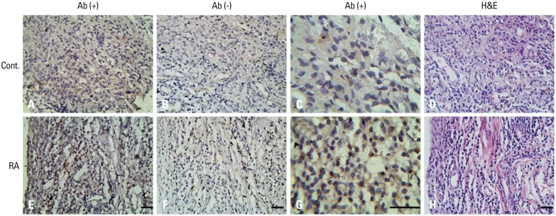
Fig. 4. Immunohistochemical study of CCL2 expression in synovial tissue of knee joints. Photomicrographs were taken from healthy controls (A-D), rheumatoid arthritis patients (E-H). (A, C, E, and G) CCL2 immuoreactivity (brown staining). (B and F) Background staining with CCL2 antibody omitted (blue staining). (H) Maximal inflammatory cell infiltration (hematoxylin and eosin staining in D and H; avidin-biotin-peroxidase staining in A-G. Original magnification ×400 in A, B, D, E, F, and H; ×1000 in C and G). Bar=40 µm.

