. 2015 Jun 5;56(4):1134–1142. doi: 10.3349/ymj.2015.56.4.1134

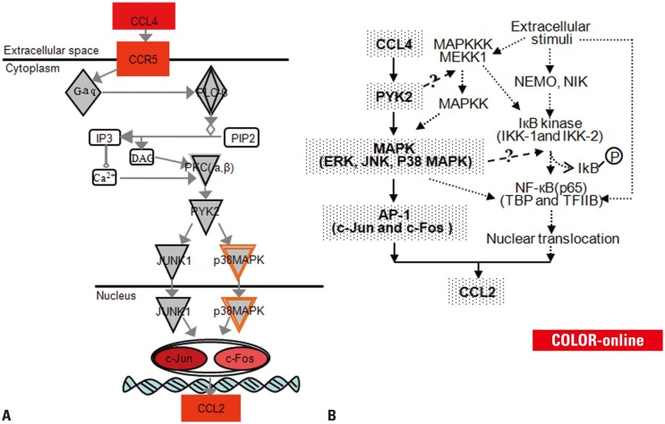
Fig. 6. (A) Schematic representation of CCL4/CCR5/c-Jun and c-Fos/CCL2 signaling pathway in RA. Red color indicates gene increased expression, the darker the higher expression. (B) Signal transduction cascades in rheumatoid arthritis and factors that stimulate the CCL2 transcription. Signaling molecules that we have confirmed are shown in coarse stippling, and confirmed pathways are shown with bold arrows. Suspected factors that may be involved are shown in white, and potential pathways are shown with dashed arrows. DAG, diacyl glycerol; PIP2, phosphatidylinositol 4,5-trisphosphate; PKC, protein kinase C; PYK2, prolinerich tyrosine kinase 2; JUNK1, Jun N-terminal kinase 1; MAPK, MAP kinase; NEMO, NF-κB essential modulator; NIK, NF-κB-inducing kinase; IKB, inhibitors of NF-κB; IKK, IκB kinases; ERK, extracellular signal-regulated kinase; JNK, Jun N-terminal kinase; AP-1, activator protein; TBP, TATA-binding protein.

Fig. 6