Figure 1.
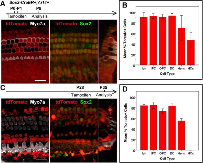
A,B. Sox2-CreER labels >85% supporting cells and >50% inner and outer hair cells in the organ of Corti when induced at P0-1. C,D. Sox2CreER labels only supporting cells in the organ of Corti when induced at P28. Wholemount cochlear confocal images (A,C) and quantification (B,D) of Sox2-CreER; ROSA-CAG-Stop-floxed-tdTomato (Ai14) when induced at P0-P1 and analyzed at P8, or induced at P28 and analyzed at P35. Hair cells are labeled with Myo7a antibody (white) in the P0-P1 induced samples (A), and in the P28 induced samples (C). Sox2 immunostaining (green) labels supporting cells, and tdTomato (red) labels Cre activity. (B and D). Percentages of tdTomato+ cells in each subtype of supporting cells (Inner phallengeal cells [Iph], Inner pillar cells [IPC], outer pillar cells [OPC], Deiters’ cells [DC], Hensen cells [Hens]) and hair cells (HCs). Scale bars = 20 μm. Error bars = ±1 standard error in three independent mice.

