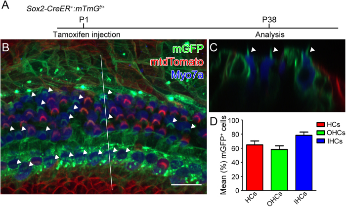
Figure 2. Sox2-CreER labels both supporting cells and hair cells when induced at P1.
Tamoxifen was intraperitoneally injected into Sox2-CreER; mTmG mice at P1 and cochleae were analyzed at P38. A. The whole mount of apical cochlear turn with mGFP (green) and mtdTomato (red) fluorescence and Myo7a immunostaining (blue) using LSM700 confocal laser scanning microscope. B. The optical xz-plane of the line in A. Arrowheads label mGFP+ hair bundles. Scale = 20 μm. C. Percentages of mGFP+ hair cells in a 300-μm region from the apical turn. Error bars = ±1 standard error, N = 3 independent mice.

