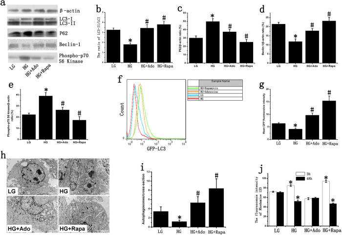
Figure 2. Effect of adenosine on late EPC autophagy.
(a) LC3, P62, Phospho-p70 S6 Kinase and Beclin-1 were determined by western blotting. We used cropped gels; the gels were run using the same experimental conditions. (b–e) The expression of LC3-I, LC3-II, P62, Beclin-1 and Phospho-p70 S6 Kinase in late EPCs. β-actin was used as a loading control. (f,g) The mean LC3-GFP fluorescence intensity of late EPCs. The mean LC3-GFP fluorescence intensity was decreased after stimulation with HG, and adenosine could increase HG-inhibited autophagy. (h) TEM confirms the presence of autophagosomes in the late EPCs. (i) Quantitative analysis of the number of autophagosomes in late EPCs. (j) The mean fluorescence intensity of rhodamine 123 in late EPCs. The cells were stained with rhodamine 123 after 3 or 48 hours of stimulation, and the average fluorescence values were analyzed using flow cytometry. The groups were as follows: low glucose (LG); high glucose (HG); high glucose + adenosine (HG+Ado); high glucose + rapamycin (HG+Rapa). *p < 0.05 (n = 4) versus LG. #p < 0.05 (n =4) versus HG. Values are the mean ± SE.

