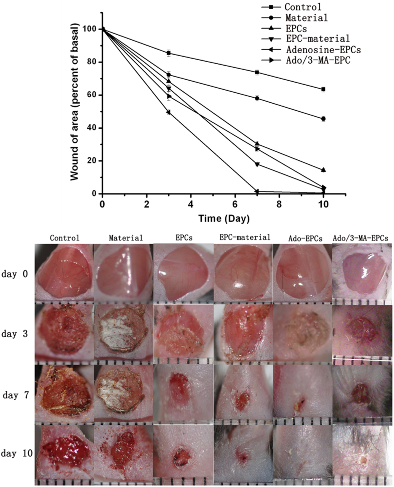
Figure 6. Stimulation of EPCs with adenosine to promote diabetic wound healing.
(a) Adenosine-stimulated EPCs effectively promoted diabetic wound healing. Compared with the control group, the biological material alone and signal EPCs displayed faster wound healing rates. The adenosine-stimulated EPCs showed significantly improved wound healing compared with the normal EPC-Biomaterial group, and autophagy inhibition could inhibit the effect of adenosine on diabetic wound closure. (b) Photographic documentation of wounds at days 3, 7 and 10. The wound was very well healed on day 7 and was completely closed on day 10 in the adenosine-stimulated EPCs group. *p < 0.05 (n = 10) versus the control group. Values are the mean ±SE; #p < 0.05 (n = 10) versus the EPC-Biomaterial group.

