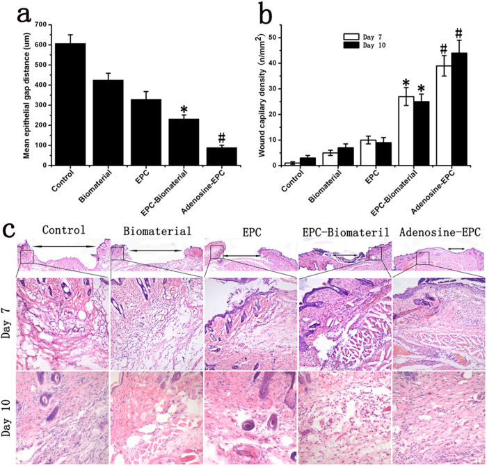
Figure 7. Effects of adenosine-stimulated EPCs on angiogenesis in diabetic wound.
(a) Mean epithelial gap distance on day 7. (b) The wounds capillary density on day 7 and day 10. The analysis of wounds capillary density was based on H&E staining. The biological material and signal EPC group promoted angiogenesis compared with the control group. The material seeded with adenosine-stimulated EPCs increased the vascular density compared with the normal EPC group. (c) We performed H&E staining to observe the epithelial gap and vascular density in each group. Wounds treated with adenosine-stimulated EPCs seeded on the biomaterial had significantly reduced epithelial gaps compared with wounds treated with EPCs seeded onto the biomaterial on day 7. The highest vascular density was found in the adenosine-stimulated EPC group. *p < 0.05 (n = 10) versus EPC; #p < 0.05 (n = 10) versus EPC-Biomaterial. Values are the mean ± SE.

