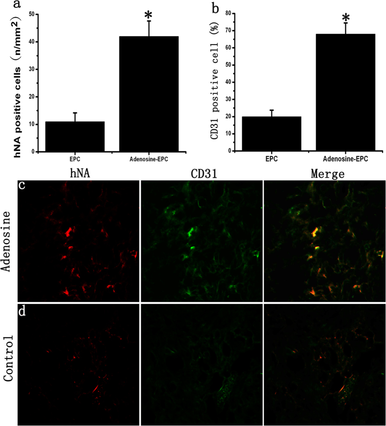
Figure 8. Effects of adenosine on the survival of transplanted EPCs in diabetic wounds.
(a) The number of EPCs remaining in the diabetic wound of the adenosine-stimulated EPC group was 4.35-fold higher than that of the unstimulated EPC group. (b) The ratio of CD31-positive cells relative to the number of EPCs remaining in the diabetic wound. In the adenosine-stimulated EPC group, the number of remaining stem cells participating in angiogenesis was greater than that in the unstimulated EPC group. (c–d) Vascular endothelial cells were stained with a CD31 antibody (green), and human-derived cells were stained with an hNA antibody (red). *p < 0.05 (n = 10) versus control. Values are the mean ± SE.

