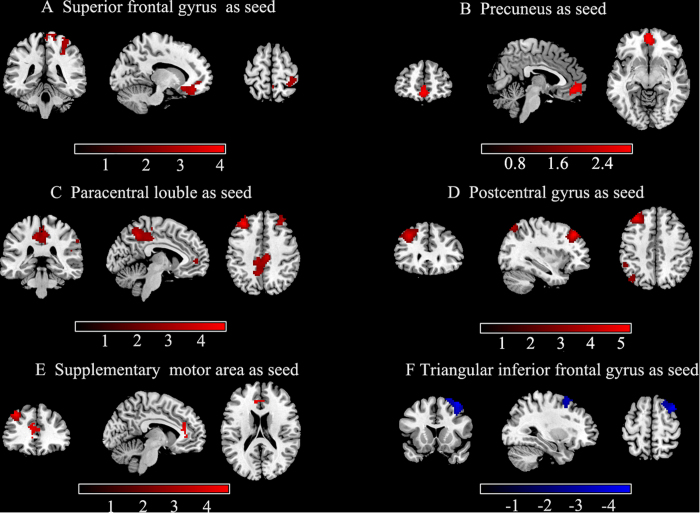
Figure 2. Group differences of inter-regional FC maps between seeds and the rest voxels of the brain.
Six abnormal fALFF regions (superior frontal gyrus, precuneus, paracentral lobule, postcentral gyrus, supplementary motor area, and inferior frontal gyrus) were defined as seeds for FC analyses. Compared to healthy controls, patients with PNES showed increased (red) and decreased (blue) connectivity based on these seeds. The results were corrected by AlphaSim(all the clusters survived p < 0.05, a combined threshold of p < 0.01 with a minimum cluster sizes of 116,115,162,150,79 and 157 voxels, respectively). Color bar indicates the T score. More details of altered FC regions are described in Table 3.

