FIG. 6. .
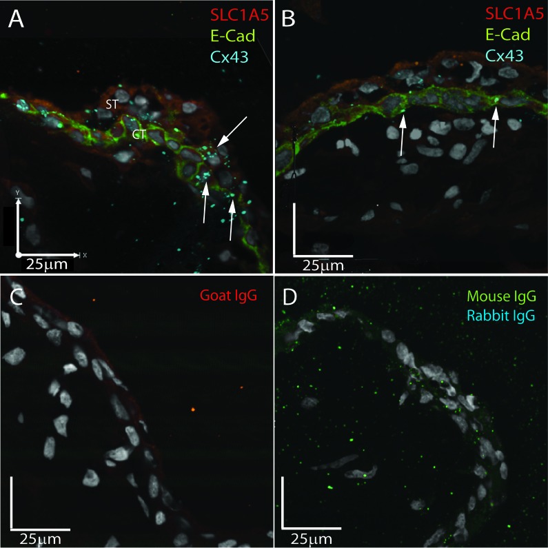
Fusing cytotrophoblast demonstrates in situ colocalization of Cx43 and SLC1A5. Representative triple immunofluorescent images of SLC1A5 (red), E-cadherin (green), and Cx43 (blue) expression in first-trimester placental sections are shown. A) In areas of cell fusion shown by loss of E-cadherin, Cx43 and SLC1A5 colocalize (arrows) to the junction of the apical membrane of the cytotrophoblast and the basal membrane of the syncytiotrophoblast. B) In quiescent areas of the same placenta, no colocalization is observed, and Cx43 is restricted to the cytotrophoblast cell junctions. C) Nonspecific goat IgG control. D) Nonspecific dual IgG controls of mouse and rabbit IgGs. Bar = 25 μM.

