
Figure 4.
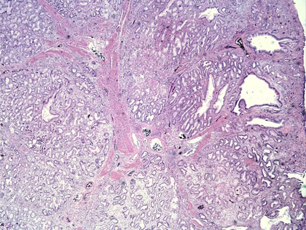
Histopathological image of polyp with admixture of fibromuscular and adipose tissue within and surrounding lobules of Brunner glands, few of which are cystically dilated (H&E; ×400).
Official websites use .gov
A
.gov website belongs to an official
government organization in the United States.
Secure .gov websites use HTTPS
A lock (
) or https:// means you've safely
connected to the .gov website. Share sensitive
information only on official, secure websites.
Histopathological image of polyp with admixture of fibromuscular and adipose tissue within and surrounding lobules of Brunner glands, few of which are cystically dilated (H&E; ×400).