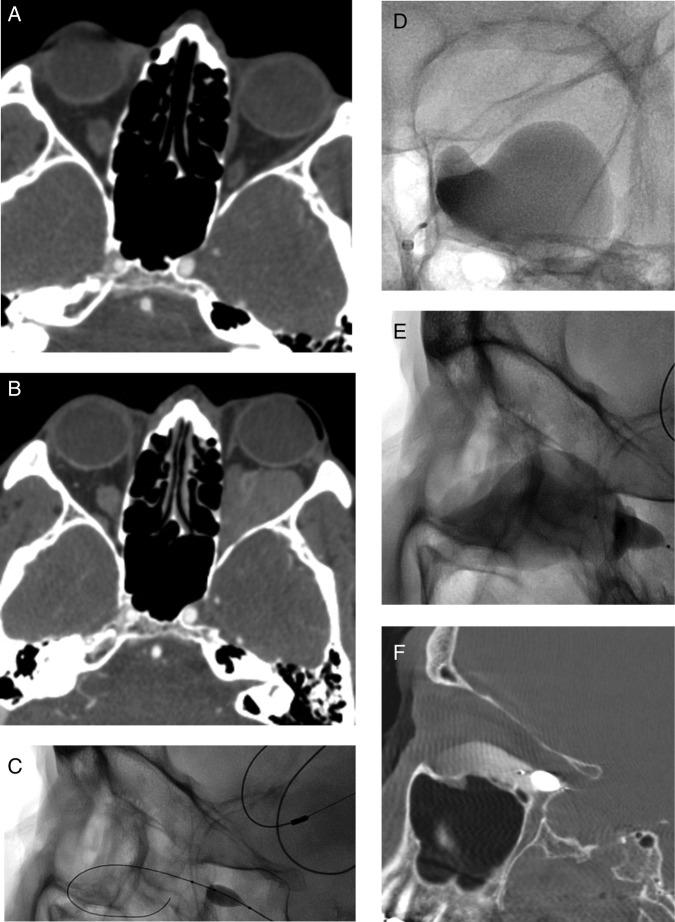
Figure 2.
(A, B) Axial contrast-enhanced CT images of the orbit demonstrate a small mass within the inferior aspect of the left orbit (A) that enlarges during the Valsalva maneuver (B). (C) Lateral spot fluoroscopic imaging shows a balloon at the origin of the left inferior orbital vein with a wire coiled in the venous varix. (D, E) Anteroposterior (D) and lateral (E) spot fluoroscopic images show a balloon at the origin of the left inferior orbital vein occluding flow into the cavernous sinus with bleomycin mixed with contrast opacifying the venous varix. (F) Sagittal dynaCT shows contrast mixed with bleomycin in the venous varix with a microcatheter balloon at the origin of the inferior ophthalmic vein.

