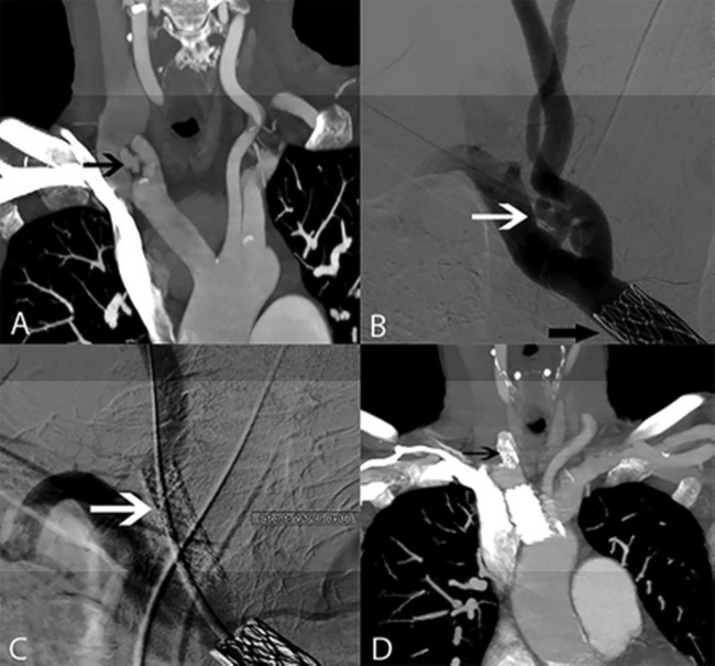
Figure 2.
(A) CT angiography (CTA) shows early contrast filling of the internal jugular vein (IJV) related to the fistula from the proximal common carotid artery (CCA) to the IJV (black arrow). (B) Digital subtraction angiography shows the fistula (white arrow) with faint IJV filling as well as the previously placed stent in the brachiocephalic artery (black arrow). A V12 covered stent (white and black arrows in C and D, respectively) is placed in the proximal CCA just from its origin to exclude the fistula (C) with disappearance of early IJV filling. On control CTA after 3 months (D), there is no evidence of residual fistula filling.

