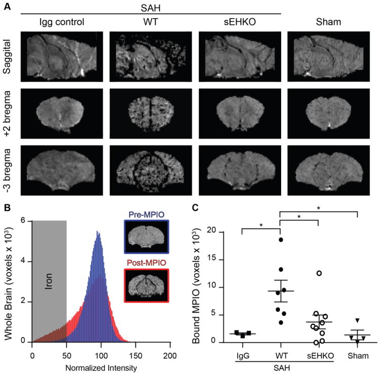
Figure 4.

sEHKO mice have less VCAM-1 expression than WT mice after SAH. A.) Representative T2* weighted images in SAH mice (IgG control, WT, and sEHKO) and sham operated mice. Deposition of the VCAM-1 MPIO causes hypointensity on MRI. B.) Representative normalized histogram of voxel intensities within a single WT brain 24h after SAH pre-MPIO (blue) and post-MPIO (red) injection (80min). Voxels below the normalized intensity threshold were used to measure the extent of iron particle deposition. C.) Quantification of MPIO deposition as the increase in iron particles detected 80min after injection in IgG control (n=3) WT SAH (n=7) sEHKO SAH (n=9) and Sham (n=4). sEHKO mice have reduced MPIO deposition compared to WT mice (*=p<0.05).
