Figure 3.

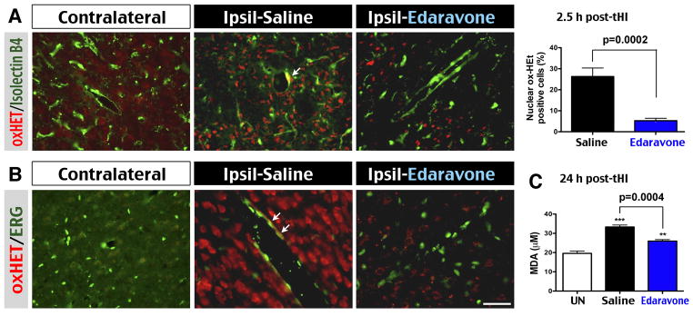
Edaravone abated oxidative stress in the brain parenchyma and vascular endothelium. A, Merged image of oxidized hydroethidine (oxHET) and isolectin B4 (an endothelial marker) labeling revealed more superoxide-positive cells in the ipsilateral hemisphere in saline-treated than Edaravone-treated mice at 2.5 h post-tHI (p=0.0002 by t-test, n=8 for each). B, Higher magnification showed frequent co-localization of oxHET in ERG+ endothelial cells (arrows) in the ipsilateral hemisphere of saline-treated mice. Note the seemingly more numerous oxHET+ nuclei due to brighter fluorescence under higher magnification in B than in A. C, Quantification of malondialdehyde (MDA) suggested significant reduction of lipid peroxidation by the Edaravone treatment at 24 h post-tHI (p=0.0004 by t-test, n=5 for each). **p<0.01, ***p<0.001 compared with UN group by unpaired t-test. Scale bar: 40 μm in A; 20 μm in B.
