
Figure 12. Fluorescence photomicrographs of an islet from a neonate with an acute coxsackievirus infection.
Viral VP1 (green; A, D) co-localizes with glucagon (red; B, D) in certain cells (orange arrows) and with insulin (light blue; C, D) in another (white arrow). Nuclei were stained with DAPI (dark blue) in the merged image (D).

Figure 12—figure supplement 1. List of human samples used.
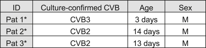
Cases were randomly selected from a previously described collection (Richardson et al., 2009).