
Figure 3. Pancreatic α cells are more resistant than β cells against CVB5- but not against cytokine- or PIC-induced cell death.
FACS-purified rat β and α cells (>90% purity for both cell types) were treated with interleukin-1β (IL-1β) + type II interferon (IFNγ) (50 or 500 U/ml, respectively) (A) or PIC (1 μg/ml) for 24 hr or infected with CVB5 (multiplicity of infection—M.O.I. 5) for 36 hr (C–G). (A–C) Apoptosis was evaluated by staining with the nuclear dies Hoechst 33342 and PI. (D) VP1 mRNA expression was assayed by RT-PCR and normalized by the housekeeping gene GAPDH. (E and F) The figures show representative Western blots of VP1 protein expression after CVB5 infection and α-tubulin for loading control. (F) The Western blot (E) was overexposed to allow visualization of VP1 expression in α cells. (G) Titration of the supernatants from β and α cells infected with CVB5 for 36 hr. Results of 4–6 experiments are plotted as box plots, indicating lower quartile, median, and higher quartile, with whiskers representing the range of the remaining data points; *p < 0.05, **p < 0.01, and ***p < 0.001 treated vs untreated (A and B) or CVB5 vs mock infection (C, D, and G); ###p < 0.001 as indicated by bars; ANOVA followed by Student's t-test with Bonferroni correction.

Figure 3—figure supplement 1. Dose-response of cytokine-induced apoptosis in pancreatic α and β cells.
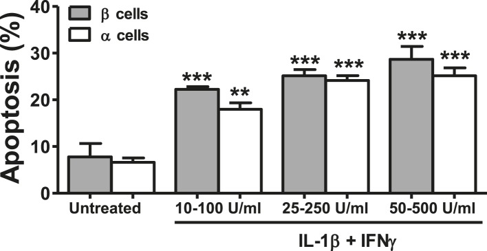
Figure 3—figure supplement 2. Pancreatic rat α and β cells have similar NO production, cytokine, and chemokine expression following exposure to cytokines.

Figure 3—figure supplement 3. Pancreatic rat α and β cells have similar cytokine and chemokine expression following exposure to PIC.

Figure 3—figure supplement 4. Prolonged time-course of CVB5-induced apoptosis in pancreatic α and β cells.
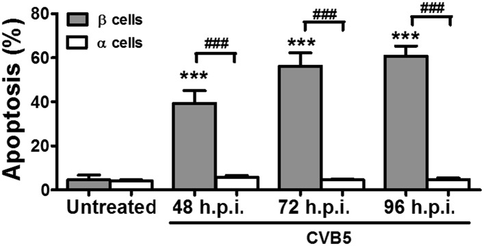
Figure 3—figure supplement 5. UV-inactivated CVB5 does not induce cell death in pancreatic α and β cells.

Figure 3—figure supplement 6. Cell counting after CVB5 infection of pancreatic α cells.

Figure 3—figure supplement 7. Pancreatic α cells infected with CVB5 under different medium conditions.
